

When Should You See a Doctor for a Breast Lump?

Dr Chong Chee Keong
Breast, Colorectal and General Surgeon
MBBS (Singapore), FRCS (Edinburgh), FRCS (Glasgow), FAMS (Singapore)
Specialist Accreditation in Intensive Care Medicine
Specialists Accreditation Board, Medical Director & Senior Consultant

Dr Chong Chee Keong Breast, Colorectal and General Surgeon MBBS (Singapore), FRCS (Edinburgh), FRCS (Glasgow), FAMS (Singapore) Specialist Accreditation in Intensive Care Medicine Specialists Accreditation Board, Medical Director & Senior Consultant
Finding a breast lump often raises an immediate question: could it be cancer? Many breast lumps are benign, including cysts and fibroadenomas, but the way a lump feels is not enough to determine its cause. A painless lump is not automatically harmless, just as a painful one is not necessarily serious.
A new breast lump should therefore be medically assessed rather than diagnosed by touch or monitored indefinitely at home. Depending on your age and clinical findings, assessment may involve breast imaging and, when necessary, a biopsy to establish what the lump represents.
Are All Breast Lumps a Cause for Concern?
Breast lumps can develop from several changes within breast tissue, and many are non-cancerous. What matters is establishing the cause rather than judging the lump by how it feels.
Common Benign Causes of Breast Lumps
Common non-cancerous causes include:
- Breast cysts: Fluid-filled lumps that may change in size or tenderness.
- Fibroadenomas: Solid benign lumps that are particularly common in younger women and often feel mobile beneath the skin.
- Fibrocystic changes: Hormone-related breast changes that can cause areas of lumpiness or tenderness.
- Fat necrosis: Firm lumps that can develop after injury or previous breast surgery.
Whether a lump is benign still needs to be established through appropriate assessment rather than its appearance or texture alone.
Can You Tell Whether a Breast Lump is Cancerous by Touch?
No. Although breast cancers are sometimes described as hard, irregular or painless, there is no reliable set of physical characteristics that can confirm whether a lump is cancerous. Benign lumps can also feel firm or irregular, while breast cancer does not always match the commonly described pattern.
This is why a new breast lump should not be self-diagnosed based on whether it moves, hurts or feels smooth. Clinical examination and appropriate imaging provide more useful information, with biopsy used when tissue diagnosis is needed.
When Should a Breast Lump Be Checked by a Doctor?
Any new breast lump should be assessed, but certain changes make timely medical review particularly important. These include a lump that persists or changes, as well as changes affecting the breast skin or nipple.
A New or Persistent Lump
A newly noticed breast lump should be medically assessed, particularly if it persists or becomes more noticeable over time. Whether the lump is small or painless does not reliably indicate what is causing it.
Any new breast lump after menopause also warrants assessment rather than being attributed to hormonal breast changes.
Changes in the Breast Skin or Nipple
A breast lump should also be assessed when it occurs alongside a new change in the skin or nipple. Relevant changes include:
- Dimpling, puckering or persistent thickening of the breast skin
- A nipple that has recently become inverted
- Spontaneous or bloody nipple discharge
- Persistent redness, scaling or other changes around the nipple
These changes can have causes other than breast cancer, but they should not be assumed to be harmless without assessment.
The Lump or Breast Is Changing
Further assessment is particularly important if a lump appears to be enlarging or the affected breast develops noticeable swelling. Redness, warmth and increasing tenderness may suggest inflammation or infection and can also require prompt treatment.
Pain alone cannot determine whether a breast lump is benign or cancerous, so a painful lump still needs to be assessed according to the overall clinical picture.
Does Family History Change How a Breast Lump Is Assessed?
A family history of breast or ovarian cancer, certain inherited genetic changes or previous breast disease may affect an individual's breast cancer risk. Tell your doctor about these factors when a new lump is assessed, as they may influence the investigations or follow-up recommended.
However, many people diagnosed with breast cancer do not have a strong family history. The absence of known risk factors is therefore not a reason to dismiss a new breast lump.
How Is the Cause of a Breast Lump Diagnosed?
Diagnosing a breast lump usually involves comparing what is found during examination with the findings on breast imaging. If these do not provide a sufficiently clear explanation for the lump, tissue sampling may be needed.
What Does the Doctor Assess During the Examination?
Your doctor will ask when you first noticed the lump and whether it has changed since then. Your menstrual history, previous breast conditions and relevant family history may also help provide context.
During the examination, the doctor assesses the lump's location and characteristics, along with the surrounding breast tissue. The nipple, skin and lymph nodes under the arm may also be examined for associated changes.
Choosing the Appropriate Breast Imaging
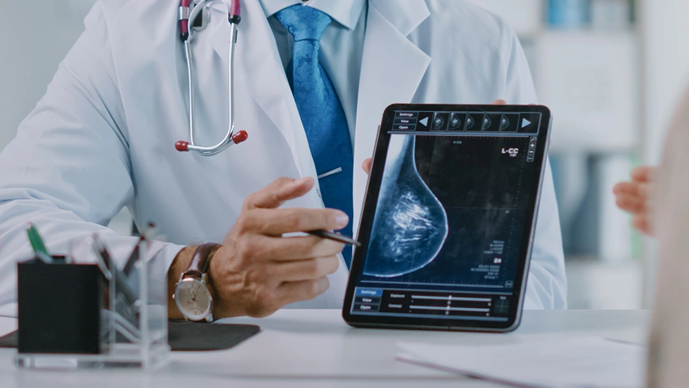
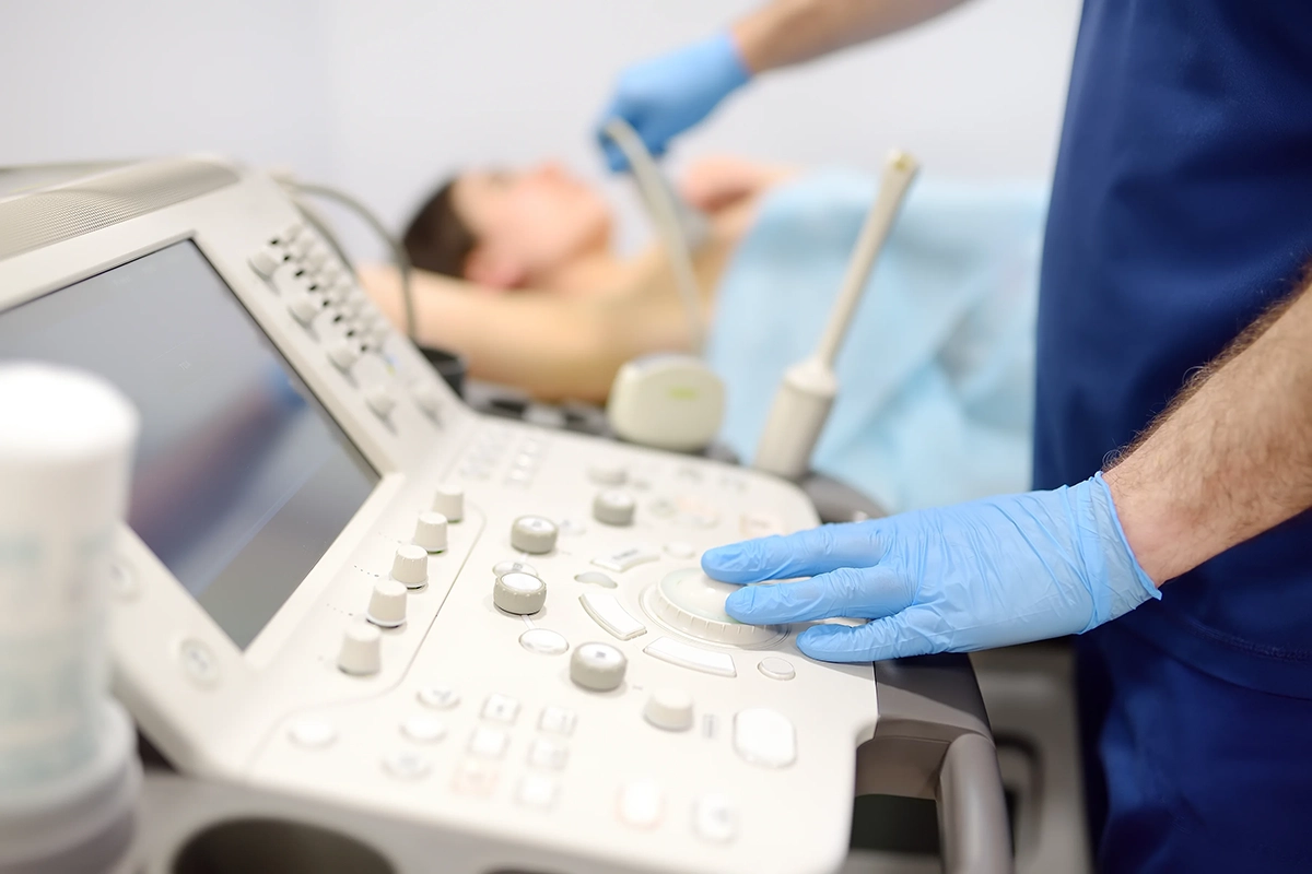
The imaging recommended depends on factors such as your age, the examination findings and the characteristics of the lump. Ultrasound can help determine whether a lump is solid or fluid-filled, while mammography provides a broader assessment of the breast tissue and can identify abnormalities that may not be apparent on examination.
Some patients may need both tests because they provide different information. MRI is generally reserved for selected situations where additional imaging is clinically useful rather than being routinely required for every breast lump.
When a Biopsy Is Needed
A biopsy may be recommended when imaging identifies a suspicious or uncertain abnormality, or when the imaging findings do not adequately explain what was found during examination.
During a needle biopsy, samples of tissue are taken from the lump and examined by a pathologist. This can establish whether the abnormality is benign or cancerous and, if cancer is identified, provide information that helps guide subsequent treatment.
Once the cause of the lump has been established, management is based on the diagnosis rather than the presence of a lump alone. Some benign lumps can be monitored, while others may require treatment because of their size, symptoms or other clinical findings. If breast cancer is diagnosed, the next steps are planned according to the type and extent of the disease.
Get a Breast Lump Assessed by Dr Chong
A breast lump may require monitoring, further investigation or treatment depending on its cause. A breast specialist can assess the clinical findings alongside appropriate imaging and determine whether a biopsy or other management is needed.
At Maven Surgery, patients can receive assessment for breast lumps and other breast concerns, with further investigations or surgical treatment arranged where clinically appropriate. Dr Chong Chee Keong is a Breast, Colorectal and General Surgeon with over 20 years of clinical experience. His experience in breast surgery allows findings to be assessed in the context of whether monitoring, further investigation or surgical management may be appropriate.
If you have noticed a new or changing breast lump, speak with Dr Chong for an assessment and advice on the appropriate next step.
Frequently Asked Questions (FAQs) About Breast Lumps
Can a breast lump disappear on its own?
Can a breast lump be monitored without treatment?
Why might I need a biopsy if my breast imaging looks normal?
When should I return to the doctor after a benign diagnosis?
Will every breast lump need to be removed surgically?
Meet Our Breast Specialist in Singapore
Dr Chong Chee Keong Breast, Colorectal and General Surgeon MBBS (Singapore), FRCS (Edinburgh), FRCS (Glasgow), FAMS (Singapore) Specialist Accreditation in Intensive Care Medicine Specialists Accreditation Board, Medical Director & Senior Consultant
Dr Chong Chee Keong is a Breast, Colorectal and General Surgeon with over 20 years of clinical experience. Dual-fellowship trained and highly accredited, he founded Maven Surgery with a commitment to elevating the standard of care for breast and colorectal cancers, the two most common cancers affecting women and men.
Dr Chong works closely with multidisciplinary teams of surgical, medical and radiation oncologists and remains actively involved in international cancer conferences to stay at the forefront of advancing techniques. He has a strong focus on minimally invasive surgery, recognising the importance of preserving self-image while maintaining treatment safety and effectiveness.
“Your Partner in Every Step of Recovery and Beyond”
For appointments and enquiries, contact us via form below or call us at (+65) 6355 5285

Mount Elizabeth Novena Specialist Centre
38 Irrawaddy Road #06-33,Singapore 329563
Parkway East Medical Centre
319 Joo Chiat Place #02-05,Singapore 427989
Farrer Park Medical Centre
1 Farrer Park Station Rd #15-13,Singapore 217562
Tel : (+65) 6355 5285
Whatsapp : (+65) 6355 5285
Operating Hours: Monday to Friday: 9:00 AM to 5:00 PM Saturday: 9:00 AM to 1:00 PM Closed on Sundays and Public Holidays
